
Mole mapping also known as total body photography, is a non-invasive skin screening method that captures high-resolution images of the entire skin surface. These images document all visible moles, freckles, and pigmented lesions, creating a detailed baseline that can be used for comparison in future assessments.
Instead of relying on memory or brief visual checks, mole mapping provides objective, repeatable documentation. This allows clinicians to detect:
This level of precision significantly improves the detection of melanoma, which often begins with changes so minor they are invisible to the untrained eye.
Skin cancer often develops quietly.
In many cases, there is no pain, no itching, and no dramatic warning sign in the early stages. What appears to be a harmless mole today may slowly evolve over time, so subtly that changes are easy to miss without systematic monitoring.
This is exactly where mole mapping becomes essential.
Mole mapping is one of the most advanced and reliable approaches for the early detection of skin cancer, including melanoma. By creating a precise visual record of the skin and monitoring it over time, mole mapping helps identify suspicious changes at a stage when treatment outcomes are significantly better.
Under the care of Dr.Ehsan Sotoudeh, mole mapping is offered as a preventive medical service, not a cosmetic assessment. The focus is on medical accuracy, patient safety, and early diagnosis based on documented evidence rather than assumptions.
Mole mapping not only helps detect and monitor changes in your skin but also guides dermatologists in deciding when a mole or skin lesion should be removed. Once a suspicious or abnormal mole is identified, your doctor can safely remove it using appropriate medical techniques such as surgical excision or laser treatment.
This precise and data-driven approach ensures that only necessary lesions are treated, helping protect both your skin’s health and appearance.


This was my first time doing mole mapping, and it was much easier than I expected.

Before mole mapping, I assumed all of my moles would need to be surgically removed to be safe. Dr. Ehsan’s assessment showed that only one mole required surgery. This process saved me from unnecessary procedures and gave me real peace of mind.

Thanks to mole mapping with Dr. Ehsan, I avoided unnecessary mole removal and potential scarring on my skin. Knowing which moles truly needed treatment made a big difference for me.

The mole mapping session was very thorough and reassuring. I finally understood which moles needed monitoring and which ones didn’t.

I felt much more at ease after the mole mapping assessment. Everything was explained clearly, without pressure or unnecessary recommendations.

Mole mapping gave me clarity about my skin instead of uncertainty. The whole process felt calm and well explained.
Melanoma is among the most aggressive forms of skin cancer, yet it is also one of the most treatable, when detected early. In its initial stages, treatment success rates are exceptionally high. Once melanoma progresses deeper into the skin or spreads, treatment becomes more complex and outcomes less predictable.
The challenge is that early melanoma is usually asymptomatic. Most patients feel perfectly healthy. There is rarely pain, bleeding, or discomfort. Often, the only sign is a slow change in an existing mole or the appearance of a new one.
Mole mapping allows these changes to be identified months or even years earlier than they would be through routine checks alone. This early insight can be life-saving.
A traditional skin check is based on visual inspection during a single appointment. While valuable, it has limitations, especially for individuals with many moles or complex skin patterns.
Mole mapping enhances standard examinations by adding:
Rather than assessing the skin as a one-time snapshot, mole mapping creates a long-term visual history of your skin health.
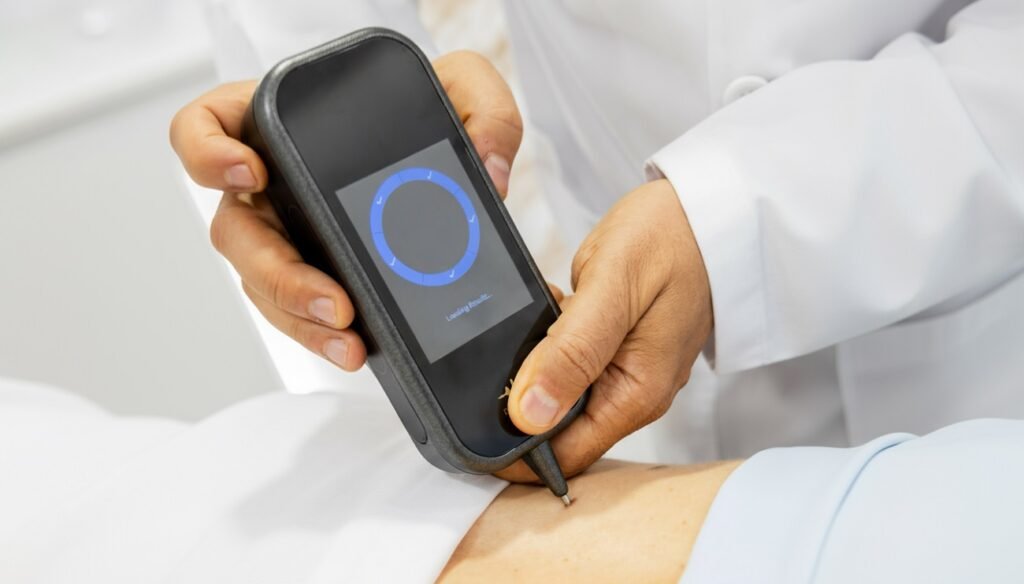
Modern mole mapping has evolved beyond photography alone. As part of advanced skin cancer screening, assessments may include AI-assisted analysis using the Dermasensor device.
DermaSensor is a handheld, non-invasive diagnostic tool that uses artificial intelligence to support the early detection of skin cancers, including melanoma, basal cell carcinoma, and squamous cell carcinoma.
The Dermasensor device analyses subsurface skin features using advanced optical technology combined with machine-learning algorithms trained on large datasets of skin lesions. Within seconds, it provides an objective risk assessment to help determine whether a lesion requires further evaluation.
Importantly, Dermasensor does not replace clinical judgment. Instead, it acts as a powerful decision-support tool that enhances diagnostic confidence, especially for lesions that appear visually ambiguous.
Early skin cancers can be difficult to distinguish from benign moles, even for experienced clinicians. AI-assisted tools help by:
When combined with mole mapping, AI-assisted assessment adds another layer of accuracy and reassurance.
Mole mapping is suitable for anyone who wants a proactive approach to skin health, but it is particularly recommended for individuals who:
Even individuals without obvious risk factors can benefit from establishing a baseline skin record for future comparison.
The appointment begins with a detailed discussion of your medical history, lifestyle factors, sun exposure, and any specific concerns you may have about your skin.
High-resolution images are taken of the entire skin surface in a systematic and consistent manner. This ensures that future images can be accurately compared.
Selected moles or lesions may be examined in greater detail using dermatoscopy or AI-assisted tools such as Dermasensor, depending on their appearance.
All images and findings are securely stored, forming your personal skin map. This record is used exclusively for ongoing monitoring and follow-up.
If any lesion requires closer attention, a personalised plan is discussed. This may involve short-term monitoring, biopsy, or referral for treatment when necessary.
The entire process is painless, non-invasive, and requires no downtime.
Not every mole change is dangerous, but certain patterns require attention. Clinicians commonly use the ABCDE framework:
Mole mapping makes it possible to track these features objectively over time, rather than relying on recollection.
Dubai’s climate involves year-round sun exposure, leading to cumulative ultraviolet damage even during routine daily activities. Many people underestimate their UV exposure because it occurs gradually rather than during intentional sunbathing.
Long-term UV exposure is one of the strongest risk factors for skin cancer. Mole mapping provides continuous monitoring that adapts to these environmental realities, making it particularly relevant for residents and long-term visitors to the region.
Yes. Mole mapping is completely non-invasive and does not involve radiation, injections, or skin damage. It is suitable for adults of all ages and can be safely repeated as part of ongoing preventive care.
The ideal frequency depends on individual risk factors:
Your follow-up schedule is tailored to your skin type, findings, and medical history.
A Smarter Alternative to Traditional Mole Biopsy
Until recently, evaluating suspicious moles often meant a surgical procedure.
In many cases, patients had to undergo minor surgery to remove a mole, send it to pathology, wait for results, and cope with discomfort, scarring, healing time, and additional costs, even when the mole ultimately turned out to be benign.
Today, this approach is no longer the first or only option.
With the advancement of AI-assisted diagnostic technology, mole assessment has become far more precise, faster, and significantly less invasive.
Using modern tools such as Dermasensor, suspicious moles can now be analysed within seconds using artificial intelligence, without cutting the skin, without stitches, and without unnecessary procedures.
Dermasensor uses advanced optical signals combined with artificial intelligence to analyse subsurface characteristics of a mole that are not visible to the naked eye. The system compares these signals against vast datasets of benign and malignant lesions, delivering an immediate clinical risk assessment.
What this means for patients:
Only moles that are truly clinically suspicious based on AI-supported analysis and medical judgment are selected for further intervention or biopsy.
This represents a fundamental shift from a “remove first, analyse later” model to a “analyse first, treat only when necessary” approach.
Surgical mole removal, even when minor, can leave permanent scars — especially on visible areas such as the face, neck, chest, or arms. For many patients, the psychological and cosmetic impact of scarring is as concerning as the procedure itself.
AI-assisted mole analysis allows clinicians to:
By limiting surgical intervention to only high-risk lesions, patients experience a safer, more comfortable, and more respectful diagnostic process.
Skin cancer is the most common cancer worldwide.
Melanoma is dangerous not because it is common, but because it spreads quickly when missed. The key difference between survival and serious disease is often how early the lesion is detected.
Melanoma develops from pigment-producing cells called melanocytes. It can appear as a new mole or arise from changes in an existing one. Unlike many other skin cancers, melanoma can metastasize rapidly if left untreated.
What makes melanoma especially concerning:
This is why technologies that detect melanoma before visible progression are so critical.
When mole mapping is combined with AI-based tools like Dermasensor, the result is a multi-layered diagnostic strategy:
This combination dramatically improves early detection while reducing unnecessary interventions.
Modern mole assessment is no longer about fear-based medicine or over-treatment.
It is about precision, prevention, and respect for the patient’s body.
By using AI-assisted analysis:
Only lesions that genuinely warrant treatment are addressedو nothing more, nothing less.
Preventive medicine is not about doing more, it is about doing what is necessary, timely, and appropriate.
AI-assisted mole analysis supports this philosophy by:
This shift represents progress not just in technology, but in how skin health is respected and managed.
Skin cancer does not need to be detected through pain, scars, or unnecessary surgery.
With modern mole mapping and AI-supported diagnostic tools, evaluation can be fast, accurate, and patient-friendly.
Only moles that truly require intervention are treated.
Everything else is monitored safely and carefully.
Early detection saves lives, and modern technology allows us to do it smarter, gentler, and more precisely than ever before.
Mole mapping is not about creating fear or leading to unnecessary procedures. It is about clarity, evidence, and informed decision-making.
By documenting the skin over time, clinicians can avoid unnecessary biopsies while ensuring that genuinely concerning changes are addressed promptly. This balanced approach protects both physical health and peace of mind.
Dr Ehsan Sotoudeh approaches mole mapping with an emphasis on:
Patients who value accuracy, transparency, and preventive care often choose mole mapping as part of their ongoing health strategy.
Skin cancer rarely announces itself loudly. More often, it appears as a quiet change, easy to overlook without the right tools and expertise.
Mole mapping, supported by modern imaging and AI-assisted assessment, offers a reliable way to identify early warning signs before they become serious conditions.
Where Mole Mapping Is Performed
Mole mapping and advanced skin cancer screening are performed by Dr.Ehsan Sotoudeh at Eden Aesthetics Clinic.
All assessments are carried out using advanced imaging and AI-assisted diagnostic tools in a professional medical setting, with a strong emphasis on accuracy, patient comfort, and evidence-based decision-making.
The goal is not unnecessary intervention, but early detection, careful monitoring, and treatment only when truly required, guided by modern technology and clinical expertise.
If you are concerned about your moles, or simply want a comprehensive and documented assessment of your skin, mole mapping offers clarity, reassurance, and early detection when it matters most.
Early detection saves lives, and it begins with careful, informed observation.
➡️ Book Your Mole Mapping Appointment